
Down syndrome 4D ultrasound—what can it actually show? Many expecting parents wonder if a 4D ultrasound can detect Down syndrome or give clear signs early in pregnancy. With advancing technology, the ability to visualize your unborn baby in such detail is both exciting and emotional—but how much can it truly reveal about chromosomal conditions?
Can you see down syndrome on 4d ultrasound? 4D ultrasounds do not diagnose Down syndrome. They provide real-time images and facial features, which may show certain markers, but a diagnosis requires genetic testing like NIPT or amniocentesis. Medical professionals use 4D scans more for bonding and surface imaging than for confirming chromosomal abnormalities.
Still, some facial indicators—like a flat nasal bridge or differences in head shape—might appear in these scans. But don’t rely on images alone. So, what should you look for, and how can parents prepare? Let’s explore expert-backed insights, diagnostic options, and what a 4D ultrasound can really tell you about your baby’s health.
Can Ultrasound Detect Down Syndrome?
Can 4d ultrasound detect down syndrome? Ultrasound is a critical tool used during pregnancy to assess the health of both the mother and the developing baby. The ability of ultrasound to detect conditions like Down syndrome, however, is a subject of ongoing research.
While a routine ultrasound cannot definitively diagnose Down syndrome, it can help identify markers or signs that may suggest the presence of this chromosomal disorder.
Ultrasound screenings typically measure the thickness of the nuchal translucency (NT) – a fluid-filled space at the back of the baby’s neck. An increased NT thickness may raise concerns and prompt further testing.
However, it’s important to note that many babies with Down syndrome do not exhibit the obvious signs during the initial ultrasound, and many babies without Down syndrome may have abnormal NT measurements.
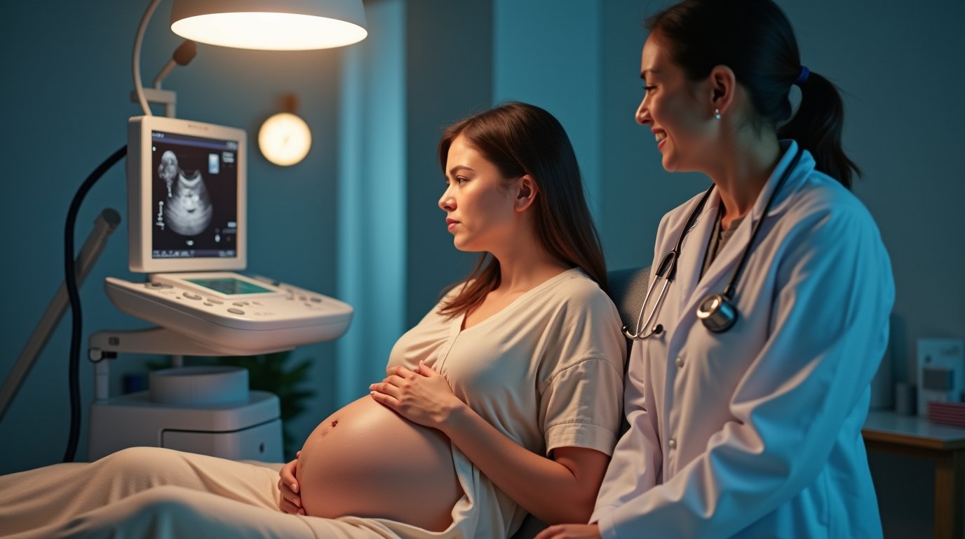
Detecting Down Syndrome on 4d Ultrasound goes a step further, providing a more detailed, real-time view of the fetus. This advanced imaging technology can offer a clearer understanding of any potential signs associated with Down syndrome and allow doctors to recommend the appropriate course of action if further testing is needed.
The Role of Ultrasound and Diagnostic Tests and Scan
While a Down Syndrome 4D ultrasound can reveal potential markers for Down syndrome, it is crucial to understand that it is not a diagnostic tool. Ultrasound results can be affected by various factors, including the baby’s position, gestational age, and maternal body type. The 4D ultrasound provides an enhanced, more detailed view compared to traditional 2D ultrasounds, but it can only reveal certain physical indicators.
Further diagnostic tests and scans are essential for confirming the presence of Down syndrome. These may include:
- Non-invasive prenatal testing (NIPT): A blood test that screens for chromosomal abnormalities, including Down syndrome. It is a highly accurate test and can be done as early as 10 weeks of pregnancy.
- Amniocentesis: This invasive procedure involves taking a sample of amniotic fluid to test for chromosomal abnormalities. It carries a slight risk of miscarriage but provides a definitive diagnosis.
- Chorionic Villus Sampling (CVS): Similar to amniocentesis, CVS involves taking a small sample from the placenta to test for genetic disorders. It can be done earlier in the pregnancy but carries a small risk of complications.
In combination with the Down Syndrome 4D ultrasound, these diagnostic tests help provide a clearer picture and can aid in making informed decisions about the pregnancy. However, the ultrasound serves as an initial screening tool that flags potential concerns, while the diagnostic tests provide confirmation.
Down Syndrome 4d Ultrasound: How Advanced Imaging Helps in Early Detection
The 4d Ultrasound Down Syndrome offers a unique advantage in the detection of Down syndrome because it combines advanced imaging technology with the ability to assess the baby’s features in real-time. Unlike traditional 2D ultrasound, the 4D version provides a more vivid and detailed view of the fetus’s face, limbs, and body structure.
Through 4D imaging, doctors can observe physical characteristics that may indicate Down syndrome. These include:
- Facial abnormalities: Babies with Down syndrome may exhibit certain facial features, such as a flattened nose bridge or upward slanting eyes. The 4D ultrasound allows healthcare providers to closely examine these features in detail.
- Heart defects: Babies with Down syndrome are more likely to have congenital heart defects. The 4D ultrasound can capture detailed images of the fetal heart, potentially detecting abnormalities early on.
- Nuchal translucency: As mentioned earlier, the 4D ultrasound provides a more detailed view of the nuchal translucency, which is a key marker for Down syndrome. A larger NT measurement can prompt further testing.
While the Down Syndrome 4D ultrasound can reveal these potential indicators, it is still only a part of the broader diagnostic process. A positive result from the ultrasound doesn’t guarantee that the baby has Down syndrome, and further tests will be necessary for confirmation.
Ultrasound Signs of Down Syndrome During Pregnancy
The Down Syndrome 4D ultrasound can help identify several key signs that may indicate the possibility of Down syndrome in the fetus. While none of these signs alone are conclusive, their presence can lead to further investigation. Some of the ultrasound signs of Down syndrome include:
- Increased nuchal translucency: A thicker NT measurement can be a sign of Down syndrome, though it can also be associated with other genetic conditions.
- Absent or hypoplastic nasal bone: In some cases, a lack of a visible nasal bone or a small nasal bone can indicate a higher likelihood of Down syndrome.
- Shortened femur length: A shorter-than-expected femur length may be another potential marker.
- Heart defects: As mentioned, structural heart abnormalities are common in babies with Down syndrome, and the 4D ultrasound can help detect these conditions early on.
It’s important to remember that Down Syndrome Ultrasound 4d cannot confirm a diagnosis. Instead, it serves as an early detection tool that allows healthcare providers to assess risk and recommend further testing if necessary. Any concerns raised during the ultrasound should be followed up with more definitive diagnostic tests, such as NIPT, amniocentesis, or CVS.
Common Questions about Detecting Down Syndrome in 4d Ultrasound (FAQs)
What is Down Syndrome?
Down syndrome, also known as trisomy 21, is a genetic condition that occurs when a person has an extra copy of chromosome 21. This additional genetic material affects physical and cognitive development, leading to various characteristics associated with the syndrome.
How can ultrasound detect Down Syndrome during pregnancy?
Ultrasound examinations can help screen for Down syndrome by identifying certain markers that may indicate the presence of the condition. Detailed ultrasound scans performed between 11-13 weeks can reveal features such as the absence of a nasal bone, which is one of the ultrasound findings associated with Down syndrome.
What are the common ultrasound findings for a fetus with Down Syndrome?
Common ultrasound findings that may suggest a risk for Down syndrome include the presence of certain markers like increased nuchal translucency, absence of a nasal bone, and specific heart defects. These features are indicative of the syndrome and may warrant further testing.
When is the best time for an ultrasound to screen for Down Syndrome?
The ideal time to screen for Down syndrome using ultrasound is during the first trimester, specifically between 11-13 weeks. However, additional ultrasounds can be conducted in the second trimester, around 20 weeks, to further assess the fetus.
What is the difference between 3D and 4D ultrasound scans?
3D ultrasound provides three-dimensional images of the fetus, allowing for more detailed visualization of anatomical structures. In contrast, 4D ultrasound adds the element of time, producing moving images. Both can be useful in assessing fetal health and detecting potential anomalies.
Can you detect Down syndrome on a 3D ultrasound?
ultrasound 3D imaging may suggest Down syndrome if physical traits (e.g., flat face, short limbs) appear, but diagnosis requires genetic testing like amniocentesis or NIPT for confirmation.
What is the role of blood tests in diagnosing Down Syndrome?
Blood tests, such as those that analyze the mother’s blood for specific markers, can also assist in screening for Down syndrome. These tests, combined with ultrasound findings, can provide a more comprehensive risk assessment for the condition.
Can Down Syndrome be confirmed through ultrasound alone?
Ultrasound alone cannot confirm Down syndrome; it can only indicate a risk. A definitive diagnosis usually requires additional diagnostic tests such as amniocentesis or chorionic villus sampling (CVS), which analyze the chromosomes of the fetus.
What are the characteristics associated with Down Syndrome?
Individuals with Down syndrome may exhibit a variety of characteristics, including distinct facial features, developmental delays, and varying degrees of intellectual disability. Early diagnosis and intervention can significantly improve outcomes for children with Down syndrome.
How does early diagnosis impact the management of Down Syndrome?
Early diagnosis through prenatal screening and ultrasound can help parents prepare for the birth of a child with Down syndrome. Understanding potential health issues and developmental challenges allows families to seek appropriate resources and support from the beginning.
What are the 4 ultrasound views of the heart?
The four standard ultrasound views of the heart are: parasternal long-axis, parasternal short-axis, apical four-chamber, and subcostal view. These views help assess cardiac structure and function.
What type of sound waves are used in ultrasound?
Ultrasound uses high-frequency sound waves (1-20 MHz) beyond human hearing. These waves bounce off tissues to create images, aiding in medical diagnostics and monitoring without radiation.
Can ultrasound tell if baby has a cleft lip?
Yes, ultrasound can often detect a cleft lip as early as 13–20 weeks, but smaller clefts may be missed. A 3D ultrasound improves accuracy. Confirmation occurs at birth.
What is the 1st, 2nd, and 3rd trimester?
The 1st trimester (weeks 1-12) involves embryo formation, the 2nd trimester (weeks 13-26) brings organ development, and the 3rd trimester (weeks 27-40) prepares trimester fetuses for birth with growth and maturation.
Conclusion
Can you detect down syndrome in a 4d ultrasound? In conclusion, the 4d Ultrasound Down Syndrome is an advanced imaging tool that can play a significant role in early detection of Down syndrome, although it is not a definitive diagnostic method. By providing a detailed view of the fetus in real-time, it helps prenatal care providers spot potential physical markers that may indicate the presence of Down syndrome. However, for a conclusive diagnosis, additional tests such as NIPT, amniocentesis, or CVS are necessary.
While the Down Syndrome 4D ultrasound is a valuable tool, it is essential for expecting parents to understand its limitations and work closely with their healthcare provider to assess risk and make informed decisions. By combining the 4D ultrasound with other diagnostic methods, parents can gain a clearer understanding of their baby’s health and be better prepared for any challenges that may arise during pregnancy.
Recommended posts
- Why Doctors Look at Palm Crease in Down Syndrome Diagnosis?
- How Long Can a Person with Down Syndrome Live? (Statistics)
- 7 Subtle Mosaic Down Syndrome Symptoms & Genetic Condition
- Down Syndrome hCG Levels by Week: Screening Risk Indicators
- 2025 ICD-10-cm Code for Down Syndrome: (Exclusive Guide)
- Can NIPT Test Be Wrong for Down Syndrome? Accuracy Explained